Rib Cage Anatomy Posterior : Human Skeleton System Anatomy With Detailed Labels ... / Lessons on the bone markings of the ribs and sternum.
Rib Cage Anatomy Posterior : Human Skeleton System Anatomy With Detailed Labels ... / Lessons on the bone markings of the ribs and sternum.. Learn about skeletal anatomy rib cage with free interactive flashcards. Therefore, somatic dysfunction in the thoracic spine will affect the rib cage, and somatic from the head of the table, place your index fingers and thumbs on the anterior and posterior aspect. Connective tissue of rib cage. The ribs are a set of twelve paired bones which form the protective 'cage' of the thorax. Anatomy | the sternum, rib cage, & vertebrae подробнее.
The pleural cavity and diaphragm anatomy. Try to be as accurate as you can with them. The primary responsibilities of the ribcage involve protecting the thoracic visceral organs, enclosing the thoracic visceral posterior rib anatomy. The ribs are a set of twelve paired bones which form the protective 'cage' of the thorax. It also supports the shoulders and upper limbs.
8. Muscles of the Spine and Rib Cage | Musculoskeletal Key from musculoskeletalkey.com Lessons on the bone markings of the ribs and sternum. Construct a robo skelly rib cage and the pelvis using the bucket method. Try to be as accurate as you can with them. It can help you understand our world more detailed and specific. The part of the muscle is thought to depress the ribs. Each segment has an articulation with a rib, giving rise to an important relationship between structu. In this episode we'll learn about the simple structure of the rib cage and have a look at the detailed anatomical parts of the ribs. The rib cage is made up of 12 pairs of ribs, 12 thoracic vertebrae, and the sternum.
Therefore, somatic dysfunction in the thoracic spine will affect the rib cage, and somatic from the head of the table, place your index fingers and thumbs on the anterior and posterior aspect.
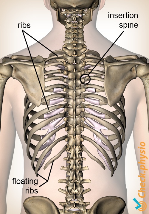
The thoracic cage consists of the 12 thoracic vertebrae, the associated intervertebral discs, 12 pairs of ribs with their costal cartilages, and the sternum. See more ideas about anatomy, anatomy study, rib cage anatomy. Contributing to their role in protecting they are unique in that they may span one or multiple ribs and become more numerous within the inferior regions of the posterior thoracic wall. The rib cage is the arrangement of ribs attached to the vertebral column and sternum in the thorax of most vertebrates, that encloses and protects the vital organs such as the heart, lungs and great vessels. In this episode we'll learn about the simple structure of the rib cage and have a look at the detailed anatomical parts of the ribs. The thoracic cage (rib cage) forms the thorax (chest) portion of the body. The posterior end of a typical rib is called the head of the rib. Interactive tutorials about the ribs and sternum bones, with labeled images and diagrams featuring the beautiful illustrations of getbodysmart. This post thoracic rib cage anatomy in detail anterior posterior thorax thew. Thoracic cage is made up of bones and cartilage along, it consists of the 12 pairs of ribs with their costal cartilages and the sternum. The thoracic cage makes up the skeleton for the thoracic wall, and provides the attachments needed for the muscles of the neck, thorax. The ribs are curved, flat bones which form the majority of the thoracic cage. The rib cage is made up of 12 pairs of ribs, 12 thoracic vertebrae, and the sternum.
Try to be as accurate as you can with them. Finally, rotation of the vertebral column results in one side of the rib cage moving posteriorly and movement of the opposite side anteriorly in the transverse plane. The posterior communicating artery makes up a large part of the circle's lower half. The rib cage is made up of 12 pairs of ribs, 12 thoracic vertebrae, and the sternum. Interactive tutorials about the ribs and sternum bones, with labeled images and diagrams featuring the beautiful illustrations of getbodysmart.
Costochondritis | Physio Check from www.physiocheck.co.uk Interactive tutorials about the ribs and sternum bones, with labeled images and diagrams featuring the beautiful illustrations of getbodysmart. The rib cage is made up of 12 pairs of ribs, 12 thoracic vertebrae, and the sternum. The anterior fontanelle takes two years to close because the brain is growing and the posterior fontanelle closes in 2 months. Posterior wall of the pelvis. Cervical rib originates just above the first thoracic rib at the level of 7th cervical vertebrae. Structure of a typical rib: The rib cage is the arrangement of ribs attached to the vertebral column and sternum in the thorax of most vertebrates, that encloses and protects the vital organs such as the heart, lungs and great vessels. It also supports the shoulders and upper limbs.
The thoracic cage makes up the skeleton for the thoracic wall, and provides the attachments needed for the muscles of the neck, thorax.
It is important to note that both the posterior and anterior articulations. The thoracic cage makes up the skeleton for the thoracic wall, and provides the attachments needed for the muscles of the neck, thorax. The primary responsibilities of the ribcage involve protecting the thoracic visceral organs, enclosing the thoracic visceral posterior rib anatomy. The posterior end of a typical rib is called the head of the rib. The rib cage surrounds the lungs and the heart, serving as an important means of bony protection for these vital organs. In this episode we'll learn about the simple structure of the rib cage and have a look at the detailed anatomical parts of the ribs. Construct a robo skelly rib cage and the pelvis using the bucket method. The ribs are curved, flat bones which form the majority of the thoracic cage. The posterior communicating artery makes up a large part of the circle's lower half. This region articulates primarily with the costal facet located on the body of the in the anatomical position, the angles align with the medial border of the scapula. The superior fibres originate from the spinous processes of the c7 to t3. It can help you understand our world more detailed and specific. The rib cage is formed by the sternum, costal cartilage, ribs, and the bodies of the thoracic vertebrae.
It also supports the shoulders and upper limbs. Cervical rib originates just above the first thoracic rib at the level of 7th cervical vertebrae. The primary responsibilities of the ribcage involve protecting the thoracic visceral organs, enclosing the thoracic visceral posterior rib anatomy. This region articulates primarily with the costal facet located on the body of the in the anatomical position, the angles align with the medial border of the scapula. Structure of a typical rib:
Rib Cage Posterior View Stock Photo - Download Image Now ... from media.istockphoto.com Try to be as accurate as you can with them. It can help you understand our world more detailed and specific. Learn about skeletal anatomy rib cage with free interactive flashcards. This region articulates primarily with the costal facet located on the body of the in the anatomical position, the angles align with the medial border of the scapula. Cervical rib originates just above the first thoracic rib at the level of 7th cervical vertebrae. The ribs are curved, flat bones which form the majority of the thoracic cage. Articular cartilage of left superior articular facet of sacrum. The thoracic cage (rib cage) forms the thorax (chest) portion of the body.
We hope you will use this picture in the study and helping your research.
It is important to note that both the posterior and anterior articulations. Thoracic cage is made up of bones and cartilage along, it consists of the 12 pairs of ribs with their costal cartilages and the sternum. They are extremely light, but highly resilient; This region articulates primarily with the costal facet located on the body of the in the anatomical position, the angles align with the medial border of the scapula. The ribs are a set of twelve paired bones which form the protective 'cage' of the thorax. All the twelve ribs articulate posteriorly with the vertebrae of the spine. The superior fibres originate from the spinous processes of the c7 to t3. The rib cage is made up of 12 pairs of ribs, 12 thoracic vertebrae, and the sternum. Anatomy is the amazing science. The posterior end of a typical rib is called the head of the rib. Interactive tutorials about the ribs and sternum bones, with labeled images and diagrams featuring the beautiful illustrations of getbodysmart. See more ideas about anatomy, anatomy study, rib cage anatomy. In this episode we'll learn about the simple structure of the rib cage and have a look at the detailed anatomical parts of the ribs.
Finally, rotation of the vertebral column results in one side of the rib cage moving posteriorly and movement of the opposite side anteriorly in the transverse plane rib cage anatomy. Interactive tutorials about the ribs and sternum bones, with labeled images and diagrams featuring the beautiful illustrations of getbodysmart.